Dr. Siwoff treats patients with the following
Vision and Neurological Complications & Disorders
What is Macular Degeneration?
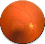
The inside of your eye has a light-sensitive tissue called the retina. The most sensitive part of the retina is a small area in the center called the macula. This area is so sensitive that if blood vessels passed through it you could see the the individual red blood cells floating through. Our eyes are wonderfully created: the macula does not get its nourishment directly from blood vessels passing through it, but rather gets nourishment from a pigment carpet and dense network of fine blood vessels under the macula. Certain people get a break or flaw in their pigment carpet. This is the dry type of macular degeneration. About 20% of these people will grow new blood vessels under their macula to get more nourishment to the area. The new vessels are fragile and tend to hemorrhage and leak. This is the wet type of macular degeneration. Although legal blindness (vision 20/200 or worse) is possible, total blindness does not occur unless there is another unrelated condition present like retinal detachment or glaucoma.
Our office has developed breakthrough technology to improve the sight of people with AMD (Macular Degeneration). For several years, we have met with success with our pioneering technique of Retinal Image Translocation (RIT), using high resolution retinal photography and precise mapping of healthy retinal tissue, as seen on the retinal fundus photos. We now use Pattern Electroretinography (PERG) to map electrical signals produced by the retina to determine the exact spot where the patient will see better. We do not require the patient’s subjective response to tell us this information, because patients with AMD typically give very inconsistent responses, depending on changing factors such as temporary health status, lighting, seating, etc. With the information from RIT and PERG, Dr. Siwoff designs custom spectacles to move the world to the exact spot of best sight in both eyes, while addressing other aspects, such as astigmatic correction and binocular vision. On average, these techniques initially result in a 200% improvement in vision. Improvement can continue to improve with the patient’s compliance with homework and doctor’s plan for followup.
What is Glaucoma?
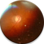
Glaucoma is a progressive disease that results in a degeneration of the nerve fibers in the optic nerve. Pressure in the eye produces this loss of vision. The eye is like a balloon. It is filled with fluid which is produced behind the iris or colored part of your eye. The fluid drains where the colored part of the eye meets the white part. This area is called the trabecula meshwork. Some people produce too much fluid. Others have a narrow angle and the fluid does not drain fast enough. In both cases, an increase in pressure in the eye damages the optic nerve which causes a loss of vision. Peripheral vision is usually the first to be affected. This is often not noticeable until it is too late. Glaucoma has been called the sneak thief of vision.
Current research with HD-OCT reveals damage to the optic nerve and retina. The primary cause of vision decline with glaucoma and optic atrophy is neurosensory loss. At Siwoff Low Vision Center, we have discovered that we are able to improve neuroconductivity by using Visual Evoked Potential (VEP) technology. The consequent result is a significant improvement in vision. For each individual with these diseases, custom spectacles are designed with precise optics necessary to uniquely improve that person’s visual function.
What is Diabetic Retinopathy?
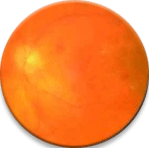
Diabetes is a disease that affects the way the body produces or processes insulin. The pancreas is the gland that produces insulin. Insulin regulates the amount of glucose in the blood. Many people think diabetes is a “sugar” disease, but all of the harmful effects of this disease result from changes in the cell walls of small blood vessels. Leaking blood vessels cause hemorrhages, exudates, new blood vessel growth, and scar tissue formation.
The standard of care for diagnosis and evaluation of the patient with diabetic retinopathy includes dilated ophthalmoscopy and fundus photography. Our effective treatment for this disease requires additional evaluative tools. Our two major breakthroughs in improving the sight of patients with diabetic retinopathy take advantage of information from HD-OCT and the PERG. These newer technologies help to locate the areas of leakage that may interfere with vision, and they also help us discover the healthiest parts of an individual’s damaged retina. The information we glean from the array of evaluative tools allows us to design spectacles that maximize the affected patient’s vision beyond what is possible with a more conventional refraction.
What is Retinitis Pigmentosa?
Retinitis Pigmentosa is an inherited progressive, degenerative eye disease that affects the rods, which are photoreceptors in the peripheral retina. Retinitis Pigmentosa causes night blindness.
For decades, electroretinograms (ERG) have been utilized to diagnose Retinitis Pigmentosa, rod/cone dystrophies, and cone dystrophies. What is new in the treatment of this disease is our discovery at Siwoff Low Vision Center that the same technology can be used therapeutically, not only to diagnose, but also to treat low vision in Retinitis Pigmentosa patients. This is a great breakthrough for people with this condition.
What is Retinal Detachment?
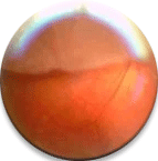
A retinal detachment can cause a total loss of sight in the affected eye. The retina is the light-sensitive tissue that is inside the eye. Trauma to the eye, high myopia, tumors of the eye, and certain diseases that produce fibrous bands of scar tissue pull the retina away from its attachment to the back of the eye.
Current Treatment:
-
Scleral buckles, cryoplexy (freezing), and lasers are used to form a scar that welds the retina back into position.
-
Retinal Image Translocation (RIT) therapy, invented by Dr. Ronald Siwoff, often helps improve vision in patients with Retinal Detachment.
Future Treatment:
-
New adhesives that are biocompatible are being developed.
-
Silicone oil, Healon and air bubbles have all been used to push the retina back into position. New materials will soon be used that will not distort vision.
Additional Disorders that we treat at Siwoff Low Vision Center
Optic Atrophy
Optic atrophy, sometimes called optic neuropathy, is a damaged optic nerve. The most common symptoms are loss of color vision, or general vision loss, in the eye that has been damaged. Since optic atrophy can take many forms and has many causes treatment is highly variable.
Uveitis
Uveitis is swelling of the middle layer of the eye, the uvea. There are a multitude of causes and factors. Uveitis is most commonly treated with medicated eye drops.
Amblyopia
Amblyopia is when one eye is not functioning as well as the other, so the brain ignores the information received by the low-function eye. Some of the most common causes are severe disorder or trauma in one eye, temporary vision loss for an extended period of time as a child or strabismus.
Strabismus
Strabismus, or heterotropia, is a misalignment of the eyes, most typically caused by trouble with the muscles around the outside of the eye. There are many possible causes, and management usually takes the form of a combination of therapy and surgery.
Keratoconus
Keratoconus is a degenerative condition which is affected by genetic and other factors. The cornea, normally dome shaped, starts thinning and sharpens into the form of a cone. This causes vision loss and blurring or duplication of images in the eye.
Rod and Cone Dystrophies
Rod or cone dystrophies are characterized by the, sometimes quite rapid, loss of rod or cone cells in the eye. This will manifest as vision loss, most notably color and peripheral vision. These disorders are often inherited.
Other Complications
Siwoff Low Vision Center is also experienced in treating patients with vision complications due to head injury, brain trauma, surgery, eye injuries and cerebral palsy, as well as many neurological disorders.


